

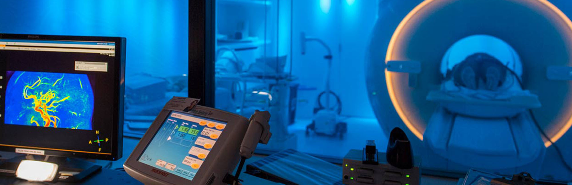
When it comes to understanding Alzheimer’s disease, standard scans are helpful, but they have limitations. By the time a radiologist can see dementia-related damage with their own eyes, the disease has usually been underway for a long time. But what if we could use advanced imaging to give patients and providers an earlier warning that dementia could be on the horizon?
My colleague, Dr. Arvind Caprihan, and I are faculty members at the Mind Research Network (part of Lovelace Biomedical Research Institute) and lead the Neuroimaging Core (NIC) of the New Mexico Alzheimer’s Disease Research Center (NM ADRC). We use advanced brain scans that are more like a complex mathematical puzzle than snapshots to catch the pre-visible signs of dementia.
Early detection reveals an opportunity for interventions that can change the course of the disease, including:
- Access to advanced treatment options and clinical trials
- Maximizing family time and access to support resources
- Cost savings for medical and long-term care
- More time to make decisions together about the future
Using advanced MRI techniques such as cerebrovascular reactivity and diffusion weighted imaging to see what the human eye can’t, we’re working with study participants to find the earliest opportunity to intervene against dementia and protect brain health.
Seeing More Than the Human Eye
The NM ADRC’s work is unique. Beyond amyloid plaques associated with Alzheimer’s disease, we focus on two factors that are very hard to see with conventional imaging:
- Inflammation of nervous tissue in the brain (neuroinflammation)
- Problems with how the brain’s small blood vessels function
Standard imaging scans may show white spots on the brain, which indicate that damage to the blood vessels has already happened. To identify small blood vessel damage before these spots appear, we use advanced neuroimaging.
Rather than recognizable photos of the brain, the raw data from these scans offer little visual information. Using advanced mathematics and computer modeling, we extract the information hidden in these scans to learn how the brain is functioning on a microscopic level before any potential damage is visible on conventional scans.
CVR: A Stress Test for the Brain’s Blood Vessels
Normally, when a patient is resting in an imaging scanner, their brain is in a restful state. This doesn’t tell us much about how the blood vessels respond to external stimuli. During a cerebrovascular reactivity (CVR) scan, participants wear a special breathing mask while inside the MRI machine. This mask delivers a small, safe dose of carbon dioxide. The gas is a vasodilator, which means it makes the blood vessels expand quickly.
By watching how the brain reacts to this tiny stressor, we can measure the ability of the blood vessels to respond when necessary, known as the vascular reserve. With this information, we can identify people whose blood vessels are struggling long before they experience memory loss or other symptoms of dementia.
Related: Read “How Blood Vessel-related Health Factors Can Influence Risk of Developing Dementia”
DWI: Tracking Water Movement in the Brain
Another powerful technique is called diffusion weighted imaging (DWI). This imaging technique allows us to track how water molecules move through the brain. In contrast to standard radiological brain images, we image motion of water molecules on a microscopic level to detect potential damage before it is visible on conventional scans.
In a healthy brain, water travels along specific tracts in the white matter that connect different areas, similar to the way cars stay in their lanes on a highway. When we see water moving around the brain in a disorganized way, it can be a powerful early warning sign of microscopic white matter damage that can’t yet be seen on a conventional scan.
This can indicate:
- Damaged brain cells’ axons
- Stroke
- Tumors
DWI is typically used to detect ischemic stroke, when a blood clot blocks an artery supplying blood to the brain.
Dedicated Space for Discovery
Advanced imaging work requires continuous access to MRI equipment, which is often in high demand at busy community hospitals. The NIC benefits from UNM Health Sciences Center’s partnership with the Center for Memory & Aging and Mind Research Network (MRN).
At MRN, our scanner is dedicated entirely to research. That means we can take time to tweak the sequences run on these scanners and develop new ways to look at the brain in a focused environment, and during daylight hours.
In addition to our work with advanced MRI, the NIC has partnerships with other centers that use PET scans to look for amyloid and tau, the specific proteins at the heart of an Alzheimer’s disease diagnosis.
Related: Read "New Mexico’s Communities Are Central to Alzheimer’s Disease Research in the U.S."
The Forefront for Future Scientists
In many labs, academic trainees and junior faculty members learn how to implement a scan developed by someone else. At the NIC, we’re at the forefront of developing the next generation of these tools.
Neuroimaging is a multidisciplinary field. It brings together people who love computer programming, mathematics, and patient care.
If you’re a trainee who thrives on solving puzzles and wants to see the direct impact of your work on a patient’s life, the NIC is the frontier. Here you’re not just studying the brain, you’re helping build the camera that could allow us to save it.
We invite you to learn more, tour our facilities, and join us as we map the future of brain health.