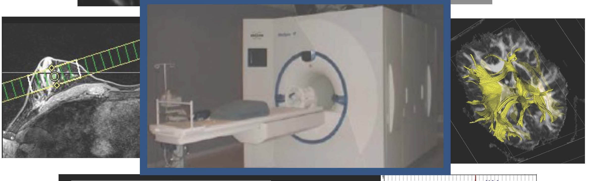
UNM School of Medicine’s Human MR Imaging Research Lab, in collaboration with scientists around the world, is breaking new ground in developing MRI techniques to help understand how the brain works and to advance diagnostic imaging in patients with cancer.
Our teams have developed advanced imaging technologies—such as real-time functional MRI—that will enable neurosurgeons to make important decisions in the operating room. These innovations push the boundaries of existing MRI technologies to explore what blood flow can tell us about brain activity.
The team’s most recent work builds on the foundations of structural and functional MRI:
- Structural MRI is a non-invasive imaging technology to map brain morphology and circulatory activity using strong magnetic fields to align nuclear spins, radio waves to change their orientation and to receive signals from nuclear spins. It can produce high-resolution images to show differences between brain tissues.
- Functional MRI (fMRI) enables doctors to measure and map brain activities in normal and diseased states by measuring changes in image intensity that are related to changes in blood flow. It can produce high-resolution images to show increases in blood flow to active areas of the brain, which produces a stronger signal in fMRI.
To develop and implement new MR technologies, our researchers work with collaborators nationally and internationally, including the University of Minnesota’s Center for Magnetic Resonance Research and the University of Copenhagen in Denmark, as well as The Mind Research Network in New Mexico.
We also train some of the brightest scientific minds in physics and engineering at the University of New Mexico and clinical scientists at the UNM Health Sciences Center. Trainees from a variety of disciplines bring new perspectives to ongoing projects that extend imaging technology with the goal to help more patients overcome neurological challenges.
TurboFIRE: Resting-state fMRI for Real-time Intraoperative Data
We are developing TurboFIRE (Turbo Functional Imaging in REal-time) to map brain activity during an ongoing fMRI scan to allow doctors to map the precise location of eloquent cortex (i.e. brain areas with specific functions) to guide brain cancer surgery.
While traditional fMRI methods map brain activity when patients engage in a task, resting-state fMRI maps functional brain systems by analyzing blood flow fluctuations when patients are at rest. By implementing imaging of the brain’s resting state in real-time using the TurboFIRE technology, we’re able to extend the benefits of MRI to injured, disabled or young patients who cannot follow the instructions to undergo traditional fMRI.
Moreover, TurboFIRE is compatible with the latest ultra-high speed fMRI data acquisition methods that provide hundreds of milliseconds temporal resolution and considerably enhance sensitivity compared with traditional fMRI data acquisition methods.
We are currently translating this technology into the intraoperative setting to allow neurosurgeons to map eloquent cortex before, during and after surgery using an intraoperative MRI scanner. Our colleagues at the University of Minnesota Medical Center are using an IMRIS scanner that is moved into the operating room to take fMRI data to monitor the success of surgery.
Using TurboFIRE, our experts can map specific functions such as language, movement or vision to their corresponding area of brain activity within millimeters while the brain is in a resting state. The objective of our current work is to determine whether resting-state fMRI can help to improve neurosurgical precision by providing a detailed map of a patient’s eloquent structures that control movement, speech and cognition.
There are many additional applications for this exciting technology, from presurgical mapping in patients for epilepsy to tracking language activation and using neurofeedback to alter brain states. TurboFIRE is currently in use at The Center for Magnetic Resonance Research and at The MIND Research Network, and we are eager to implement it at UNM Health.
High-speed Metabolic Imaging: PEPSI and Beyond
Another exciting MRI development in our lab is a high-speed MR spectroscopic imaging technology called PEPSI (Proton Echo Planar Spectroscopic Imaging) that allows us to map brain biochemicals in 3 dimensions in short scan times. MR spectroscopy is a biochemical analytic method that measures the biochemical composition of tissue based on the spectral pattern of individual biochemicals.
Spectroscopic imaging can detect biochemical changes in the brain that are associated with tumors, and these are specific for different tumor types. In other words, PEPSI allows us to rapidly take 3D, biochemical images of the brain that help doctors diagnose, manage and monitor the precise size, location and types of brain cancers.
We’ve developed this high-speed technique to enable more precise, rapid scanning. Whereas producing a typical spectroscopic image can take 20 minutes or longer, PEPSI allows us to produce 3D images in as short as three minutes. This short scan time permits us to integrate PEPSI into a clinical imaging protocol.
The future of brain imaging is wide open. More examples of research underway in our lab include:
- High frequency resting state connectivity in high-speed fMRI to overcome technical limitations of traditional resting-state fMRI.
- Mapping the diffusion properties of brain biochemical to probe the intracellular environments of brain tumors.
- Combining functional and metabolic MRI methods to simultaneously map eloquent cortex and brain biochemistry.
- Monitoring Neoadjuvant Chemotherapy Response in Breast Cancer using High-Speed 3D MR Spectroscopic Imaging of total Choline.
- Developing neurofeedback methods based on real-time resting-state fMRI to map and alter brain states.
In the Human MR Imaging Research Lab, we push the boundaries of technology every day. That’s where the exciting science happens, as we expand our knowledge of the brain, build the foundation for future advances in healthcare and extend the frontier of human knowledge.
Trainees make important contributions in the lab
While understanding the brain is serious business, our creative trainees have a lot of fun in the lab. They come from a wide range of disciplines, such as nuclear engineering, electrical engineering, physics, psychology and medicine and bring a new perspective to every project.
Their contributions are tangible. We apply a rigorous scientific approach, and often their remarkable ideas become part of our projects. Our interns and trainees not only get hands-on experience with leading-edge technologies, they also help build them.
As scientists, it’s important that we acknowledge that there is a limit to what we know. In the Human MR Imaging Research Lab, we push the boundaries of technology every day. That’s where the exciting science happens, as we expand our knowledge of the brain, build the foundation for future advances in healthcare, and extend the frontier of human knowledge.