The brain controls the body’s important functions, keeping us alive, allowing us to interact with the world and producing the thoughts and opinions that make us human.
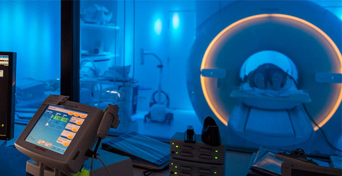
When a tumor invades, the neurosurgeon must intervene to remove the cancer as completely as possible without cutting away parts of the brain that control these important functions. Surgeons use magnetic resonance imaging (MRI) to guide them. While MRI is part of the current standard of care, there are significant barriers for precise guidance of cancer resection—creating an urgent need to enhance radiological visualization of tumor margins and infiltration and to improve efficiency of mapping brain function.
UNM Health Sciences Center has recently patented a new MRI data acquisition and analysis technology to provide surgeons with precise, real-time information about tumors and surrounding brain tissue. The technology is compatible with existing MRI scanners and performs two types of MRI scans at the same time, producing impactful data in as little as three minutes.
For the more than 1.3 million people in the U.S. living with a brain tumor, this paradigm-shifting technology could help improve outcomes after surgery.
Improving MRI Efficiency and Precision
MRI scanning relies on the ubiquity of hydrogen in living tissue (water - H 2O - is the most abundant molecule in the body), a strong magnetic field that polarizes the spin of the proton in the hydrogen atom and a radio transmitter to create a proton spin resonance that causes the protons in living tissue to emit radio signals we can process into an image.
Neurosurgeons use those images to collect information about the brain before, during and after surgery. Some of the primary types of MRI include:
- Structural MRI: Signals from different brain tissue compartments, such as gray matter in cortex and in the underlying white matter, that can be processed into a high-resolution image that reveals different brain tissue structures with high contrast.
- Functional MRI (fMRI): Highlights metabolically active tissues by detecting increased blood flow to these areas when brain activity increases, which can be processed into maps of brain functional areas. Since there is always brain activity, even when the brain is resting, the signal fluctuations in the brain can also be processed into maps of brain functional networks (resting-state fMRI).
- Diffusion MRI (dMRI): Measures water diffusivity and allows signals to be processed into maps of white matter fiber tracts that connect different cortical areas.
- MR spectroscopic imaging (MRSI): Measures signals from a multitude of intracellular organic molecules (metabolites) other than water that are specific to different tissue types and that provide information about brain biochemistry. These metabolite signals can be processed into spectral patterns and concentration maps of the metabolites. The concentrations of these molecules are changed in brain tumors and can help identify tumor types.
Functional MRI and MR spectroscopic imaging can be used to guide surgeons as they remove cancerous tissue, but collecting these MRI data is very time consuming and usually done separately in multiple sessions. MR spectroscopic imaging is technically very demanding and thus not routinely performed in clinical practice.
Moreover, task-based fMRI and intra-operative mapping, which are the current clinical standard of care and which requires patients to perform tasks to activate brain areas, have their own challenges. Patients may not be able to perform tasks due to the presence of the brain tumor, their age, or other reasons. Intra-operative mapping of language function requires patients to be awake during surgery, performing activities to engage portions of their brain so these areas can be mapped—this can be stressful for patients. This is where resting-state fMRI can be used as an adjunct to obtain data in patients who cannot perform tasks well enough to be clinically useful for surgical guidance.
Our technology provides a new high-speed approach to map brain functional networks and brain biochemistry in one imaging session, gathering hundreds of different resting state representations of whole brain function and multi-slice metabolite maps within scan times as short as 3 minutes. This opens a range of opportunities to enhance surgical removal of brain tumors while sparing eloquent cortex, in particular in the frontal cortex, which cannot adequately be imaged with conventional, task-based fMRI.
Neurosurgeons have long been focused on regions of the brain that control motor, language, visual, and auditory systems, leaving the frontal cortex largely unmapped. This technology could revolutionize how neurosurgeons make decisions, in part by providing new understanding of the brain’s frontal cortex, an important center of cognition.
The ability to collect both functional and metabolic information simultaneously could provide neurosurgeons with a new level of precision to delineate spatial margins between brain tumors and adjacent eloquent cortex to maximize tumor resection while preserving important brain function.
Related reading: Pushing the frontiers of brain imaging with real-time fMRI and more
Collaborative Research to Improve Patient Outcomes
The UNM HSC Department of Neurology has received Small Business Technology Transfer grant funding from the National Institutes of Health in collaboration with our partners at the University of Minnesota and the University of Pittsburgh to develop real-time analysis of brain activity in the presurgical setting using widely available MRI scanners and in the intra-operative setting using intra-operative MRI scanners.
This technology will be validated for presurgical mapping and intra-operative mapping in patients with brain tumors to assess the feasibility of providing neurosurgeons with planning information about brain tissue function near a tumor and with intra-operative updates of tissue function during surgery.
The new simultaneous fMRI and MRSI technology will be further developed to enable real-time resting-state fMRI mapping of eloquent cortex and online mapping of brain metabolites that are present in tissue such as amino acids, lipids, choline, creatine, and others. Comparing the quantity of these metabolites to normal brain tissue can help determine tissue metabolic status and brain tumor type.
We’re working to make these technologies widely available for clinical application, so they can have the greatest benefit for the largest number of patients. Our research involves improving data acquisition techniques to answer important clinical questions. However, this type of groundbreaking research cannot be accomplished in a vacuum, so we collaborate with partners around the nation and across the world.
UNM HSC trainees benefit from this opportunity to develop the communication and teamwork skills they need to work closely with scientists at leading research centers around the world. What’s more, they discover the entrepreneurial spirit necessary to make game-changing discoveries and publish joint papers with leading scientists worldwide.
For all of us, our foremost concern is always improving patient outcomes in the real-world setting. Our work is to make a difference in both care and surgical outcomes for patients.
Exploring your neurology education options? Request an appointment with the enrollment team. Email us today.
To find out whether you or a loved one might benefit from neurology care, call 505-272-4866.